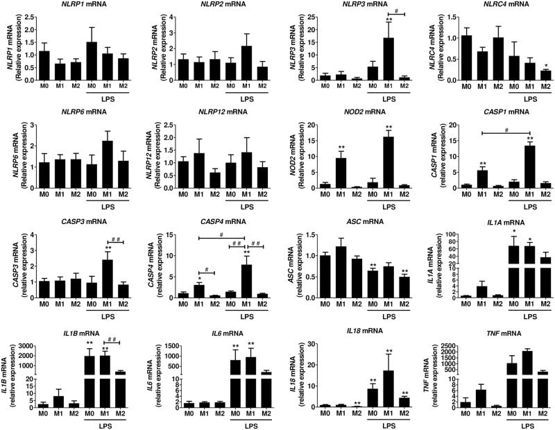
Fig 5. NLR, CASP and cytokine gene expression in human polarized macrophages.
Monocyte derived macrophages were polarized towards M1, M2, or M0 and stimulated with 100 ng/ml LPS for 3h as described in the methods. mRNA was isolated and gene expression was measured by RT-qPCR and expressed as relative fold change of M0. Data represent the mean ± SEM of ≥ 4 experiments performed in duplicates in cells isolated from independent donors. M0: cells treated with complete medium (control); M1: cells treated with 100 ng/ml IFN-γ (polarized towards M1); M2: cells treated with 10 ng/ml IL-4+IL-13 (polarized towards M2). Asterisks indicate significant differences as compared to M0 (Mann Whitney test: * p < 0.05, ** p < 0.01); (#) points out significant differences between the indicated groups (Mann Whitney test: # p < 0.05; ## p < 0.01).

