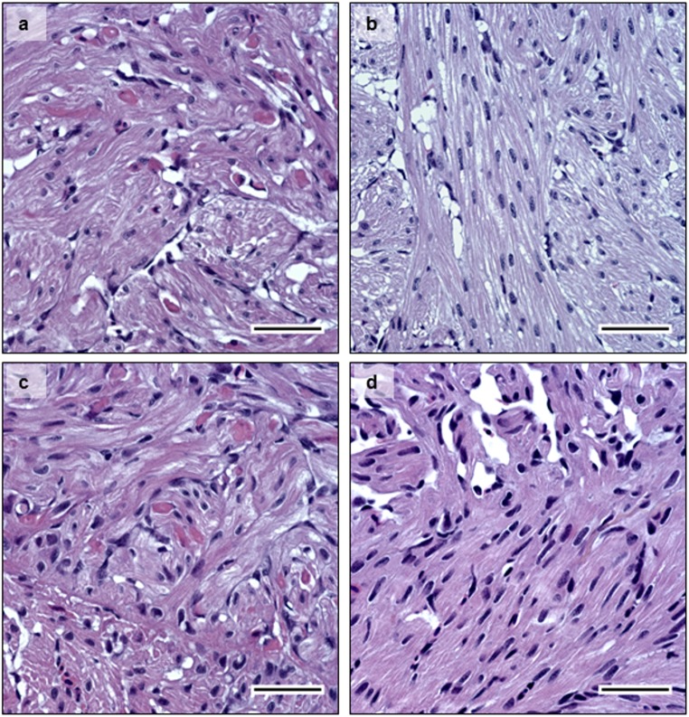
Fig 4. Histology.
HES staining of pancreas tissue sections at 14 dpe. 4a- infected diploid, 4c- infected triploid, showing cell degeneration. 4c- non-infected diploid; and 4d- non-infected triploid. Infected fish showed loss of exocrine pancreatic cells and immune cell infiltration, while non-infected controls show normal histology in pancreas. Bar = 50μm.

