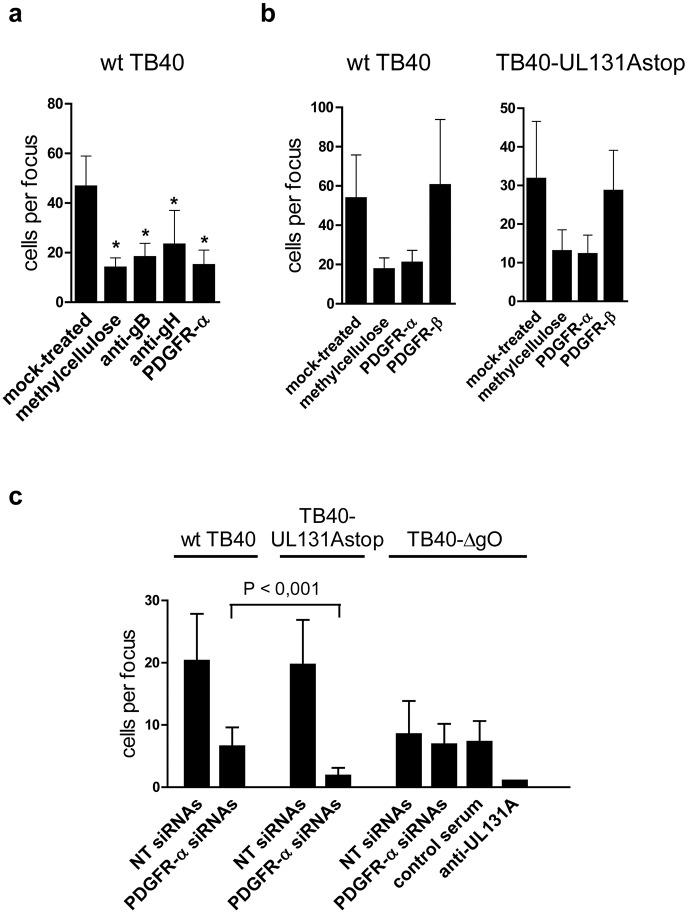
Fig 6. Silencing of PDGFR-α reduces cell-associated spread of gH/gL/gO-positive HCMV.
(a) Confluent monolayers of HFF were infected with wt TB40 virus at a very low m.o.i. After infection, cells were either overlaid with methylcellulose or medium containing anti-gB antibodies (SM5-1, 2μg ml-1), anti-gH antibodies (14-4B), PDGFR-α-Fc (2 μg ml-1), or no inhibitor (mock-treated). (b) HFF infected with wt TB40 or TB40-UL131Astop virus were mixed with uninfected cells. After adherence, cells were either overlaid with methylcellulose or fresh medium was added containing 2 μg ml-1 PDGFR-α-Fc, 2 μg ml-1 PDGFR-β-Fc, or no inhibitor (mock-treated). (a,b) 5 days later, cells were stained for HCMV IE1 by indirect immunofluorescence and cells per focus counted. For each treatment, at least 10 (a) or 20 (b) foci were counted and depicted as means +/- SD. Shown are representative experiments. Asterisks under (a) represent P<0.001 values determined by comparing foci in mock-treated monolayers with foci in monolayers overlaid with methylcellulose, co-incubated with antibodies, or co-incubated with PDGFR-α-Fc (Mann-Whitney Rank Sum test). (c) NT siRNA or PDGFR-α siRNA-transfected HFF 48 hours after transfection were mixed with HFF infected with wt TB40, TB40-UL131Astop, or TB40-ΔgO virus. After adherence, cells were overlaid with methylcellulose or methylcellulose containing anti-UL131A rabbit antiserum (1:40) or a control rabbit antiserum (1:40). 5 days later, cells were analyzed as described under (a). The P value shown (Mann-Whitney Rank Sum test) was determined by comparing cell spread of wt TB40 virus with spread of TB40-UL131Astop virus in PDGFR-α-silenced HFF.

