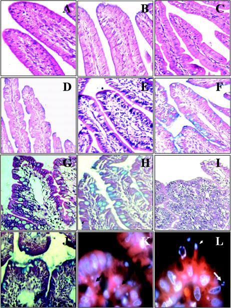
FIG. 2.
Micrographs of rabbit distal ileum tissue sections at 6 days postinfection. Intestinal sections from a PBS-treated rabbit (A and B), from an HB101-infected rabbit (C and D), from an E22ΔespA-infected rabbit (E and F), from an E22Δeae-infected rabbit (G and H), and from an E22-infected rabbit (I, J, K, and L) are shown. Sections were stained with H&E (A, C, E, G, and I), Alcian Blue (B, D, F, H, and J), or rhodamine-phalloidin and DAPI (K and L). Magnifications: A to F, ×20; G, H, and J, ×40; I, ×10; and K and L, ×100. Arrows indicate microcolonies, and the arrowhead indicates a pedestal.

