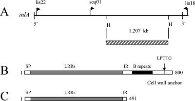
FIG. 1.
Schematic map of inlA. (A) Primers seq01 (5′-AATCTAGCACCACGTTCGGG-3′) and lis18 (5′-TCTCCTTGATTCTAG-3′) were used for specific inlA PCR amplification. The amplified DNA fragment digested with HindIII restriction enzyme (H) is represented by a hatched rectangle and cloned into the pORI19 plasmid. Structural organization of internalin A for isolates Scott A (B) and H1 (C) is shown. SP, signal peptide.

