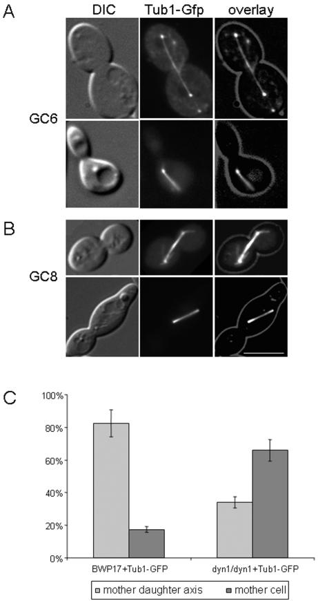
FIG.5.
Orientation of mitotic spindles in C. albicans. Spindle positions in strains GC6 (DYN1/DYN1 TUB1/TUB1-GFP) and GC8 (dyn1/dyn1 TUB1/TUB1-GFP) were analyzed by fluorescence microscopy. (A) Representative images of wild-type spindle positions in which the spindle is either aligned in the mother daughter axis and extends into the daughter (top row) or is elongated in the mother cell (bottom row). (B) Images of the spindle positions in dyn1 cells in which the spindle is either aligned in the mother daughter axis or is misaligned and elongated only in the mother cell. DIC images of cells were merged into an overlay with the images showing the GFP fluorescence. Bar, 10 μm. (C) Quantification (bars indicate the standard deviation of the mean) of spindle positioning in GC6 and GC8 (n = 130 for each strain) corresponding to the observed spindle positions in panels A and B.

