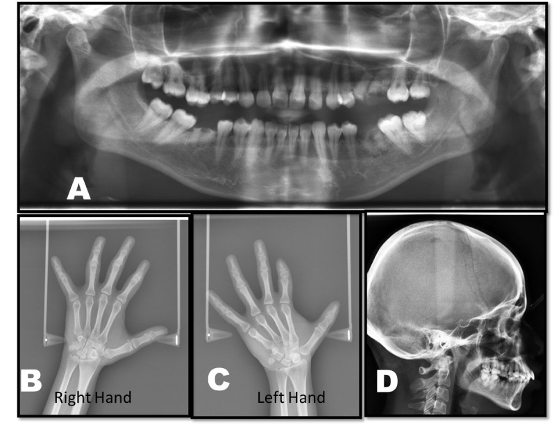
Figure 2.

Panoramic view showing decayed tooth with respect to maxillary left second premolar, first molar and mandibular right and left first molar (A). Hand wrist radiograph showing short middle phalanges in the index finger of both hands (B and C). Lateral cephalogram shows no abnormality (D).
