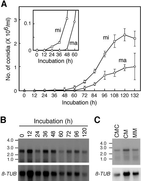
FIG. 3.
Time course of conidial development and FoSTUA expression. (A) Time course of conidial development. Mycelia of strain Mel02010 grown in CM were inoculated into CMC and incubated at 25°C for 132 h. The numbers of macroconidia (ma) and microconidia (mi) were counted at 12-h intervals with a microscope; data represent the means and standard deviations of five replications. The inset shows the time course of conidial development during the initial 60 h of incubation on an expanded scale. (B) Time course of FoSTUA expression during conidiation. Fungal tissues of strain Mel02010 grown for the indicated periods were collected, and poly(A)+ RNA was prepared from the tissues. Poly(A)+ RNA (∼5 μg/lane) was electrophoresed in a 1.5% agarose gel containing 2.2 M formaldehyde. The blot was probed with pS1BS containing the FoSTUA fragment (Fig. 2A). Sizes (in kilobases) of marker RNA fragments (Novagen) are indicated on the left. The blot was also probed with pFOTUB1 containing the β-tubulin gene fragment (β-TUB) (42). (C) FoSTUA expression during conidiation and vegetative growth. Strain Mel02010 was grown in CMC, CM, and MM at 25°C for 4 days, and poly(A)+ RNA was prepared from the cultures. The RNA gel blot was probed with pS1BS and pFOTUB1.

