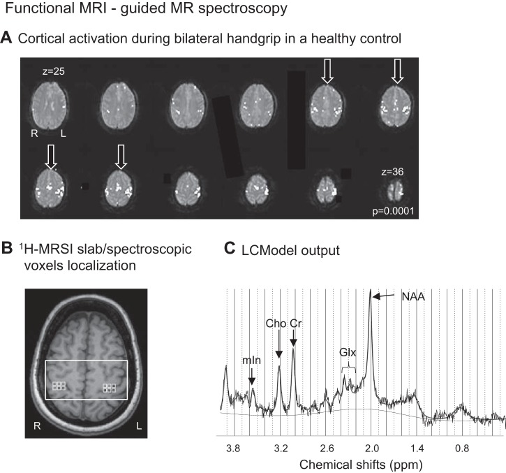
Fig. 1.
A: motor-related cortical activation during a handgrip task executed with both hands in a healthy control. The arrows indicate the four anatomical slices used to select the corresponding coincident T1-weighted image on which the 1H-MRS imaging slab was centered. R, right; L, left. B: 1H-MRSI slab (white rectangle) and MRS voxels (light gray squares) were positioned on axial T1-weighted MR image on the basis of the anatomical landmarks of the sensorimotor hand territory. C: LCModel output from one MRS voxel located in left sensorimotor cortex in a healthy control shows distinct peaks corresponding to NAA (at 2.02 ppm), Glx (2.05–2.50 ppm), Cr (3.02 ppm), Cho (3.22 ppm), and mIn (3.56 ppm) and a signal-to-noise ratio of 16.

