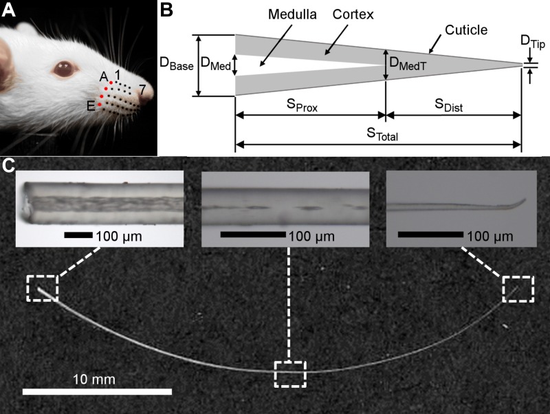
Fig. 1.
Geometric parameters of the whisker. A: each whisker is identified by its row and column position. In this photo, black and red dots have been drawn on the basepoint locations of the whiskers for visual clarity. From dorsal to ventral, the five rows are identified by the letters A–E, while the arcs (columns) are given the numbers 1–7 from caudal to rostral. The four whiskers of the caudal-most arc (colored red) are called the “Greek arc” or “Greek column” and are denoted from dorsal to ventral by the letters α, β, γ, and δ. B: schematic (not to scale) of the structural elements of the whisker. The whisker is modeled as a straight, tapered, conical frustum with base diameter (DBase) and tip diameter (DTip). The medulla is represented as a hollow cone that occupies the proximal region of the whisker. The diameter of the medulla at its base is denoted by DMed, and the diameter of the whisker at the point of medulla termination is labeled as DMedT. The total whisker arc length (STotal) is divided into a proximal portion occupied by the medulla (SProx) and a distal portion that does not include the medulla (SDist). The geometry of the cuticle was not quantified separately from the cortex in the present work. C: scanned image of the E2 whisker is shown along with three representative images to illustrate typical whisker geometry near the base, middle, and tip. The three images were not obtained from the E2 whisker; they were chosen to best illustrate the features described. The image near the base is from a C1 whisker (×10 magnification; left). The image at the location of medulla termination is from an E5 whisker (×20 magnification; middle). The image near the tip is from an E3 whisker (×20 magnification; right).

