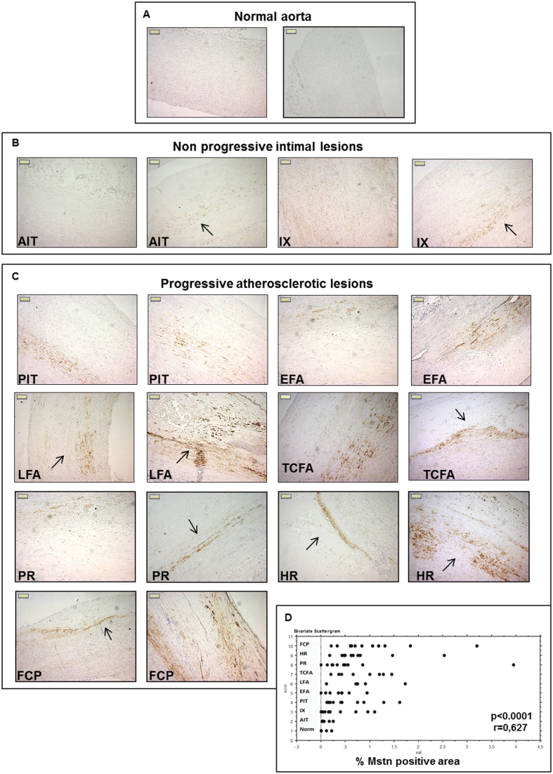
Figure 1. Expression of Myostatin (Mstn) in human peri-renal aortic specimens evaluated by immunohistochemistry.
Representative pictures of normal aortas (A), non-progressive intimal lesions (B) and progressive atherosclerotic lesions (C). (Magnification: x100; Bars = 50 μM). The arrows indicate positive areas; (D) Image analysis of Mstn immunopositivity plotted in relation to the type of atherosclerosis (Norm: Normal aorta; AIT: adaptive intimal thickening; IX: Intimal xanthoma; PIT: pathological intimal thickening; EFA: Early fibroatheroma; LFA: late fibroatheroma; TCFA: thin cap fibroatheroma; PR: plaque rupture; HR: healed plaque rupture; FCP: fibrotic calcified plaque). Mstn expression significantly correlates with lesion type progression (R = 0.627, p = 0.0001).

