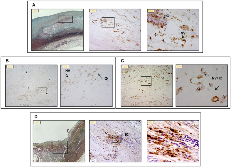
Figure 2. Myostatin (Mstn) immunostaining in progressive atherosclerotic lesions.
Panel A: late fibroatheroma, LFA; panel B: thin cap fibroatheroma, TCFA; panel C: plaque rupture, PR; panel D: healed plaque rupture, HR. Boxes indicate segments with Mstn positivity in correspondence of neovasa (NV), macrophages (Φ) and infiltrating cells (IC); magnification: 4-100x. Panel A and D: the first left image is stained with Movat pentachrome.

