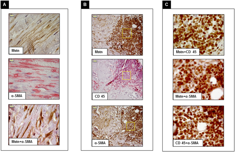
Figure 3. Immunohistochemical double staining of aortic cellular components associated with Myostatin (Mstn).
Panel A: Mstn (brown) and α smooth muscle actin (α-SMA) (red); merged image shows that Mstn colocalizes with α-SMA. Panel B: staining with Mstn (brown), CD45 (red) and α-SMA (brown). Panel C: merged images of Mstn and CD45, Mstn and α-SMA (Mstn brown; CD45 and α-SMA red) and CD45 and α-SMA (brown and red respectively), Mstn colocalizes with CD45 and α-SMA and the latter with CD45; fields correspond to boxes in panel B (Magnification: 100x panel A,C; 20x panel B).

