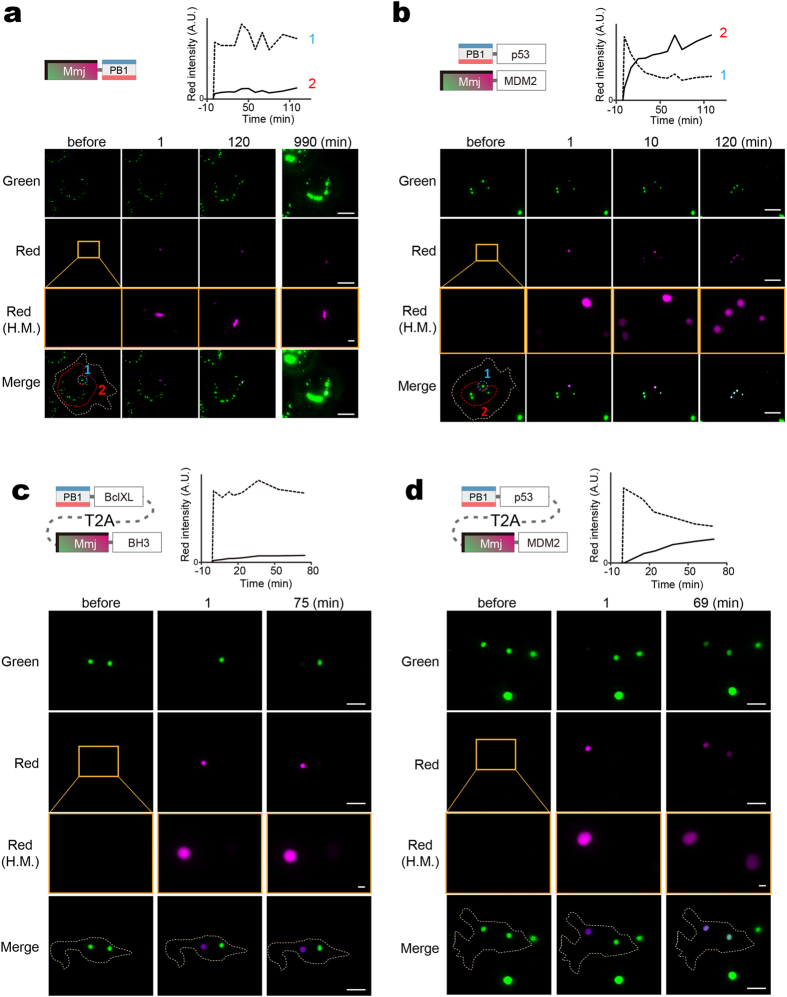
Figure 4. PcFluoppi using PB1 and Mmj for analysis of PPI kinetics.
Hydrophobic interfaces between Mmj subunits are indicated by thick bars on two adjacent sides. (a,b) Inter-punctum exchange of Mmj fluorescence in Cos-7 cells (outlined by white dotted lines) after local UV light irradiation. The photoconverted (red) fluorescence was measured in the UV-irradiated regions (1, encircled by blue dotted lines) and the intact regions (2, encircled by red dotted lines), and their intensities were plotted against time (top). (a) One day post-transfection of Mmj-PB1, cells were imaged for green and red fluorescence before and 1, 120, and 990 min after photoconversion. (b) One day post-cotransfection of PB1-p53 and Mmj-MDM2, cells were imaged for green and red fluorescence before and 1, 10, and 120 min after photoconversion. (c,d) Inter-punctum exchange of Mmj fluorescence in HEK293 cells (outlined by white dotted lines) that expressed PB1-BclXL:T2A:Mmj-BH3 (c) and PB1-p53:T2A:Mmj-MDM2 (d). In each experiment, a cell carrying two big puncta was chosen and one punctum was specifically UV irradiated. The intensities of red fluorescence from the UV-irradiated (dotted line) and intact (solid line) puncta were plotted against time (top). (a–d) Similar results were obtained from 3 other cells for each transfection. Scale bars, 10 μm. Scale bars in high-magnification (H.M.) boxes, 1 μm. See also Supplementary Fig. 4.

