
Figure 5.
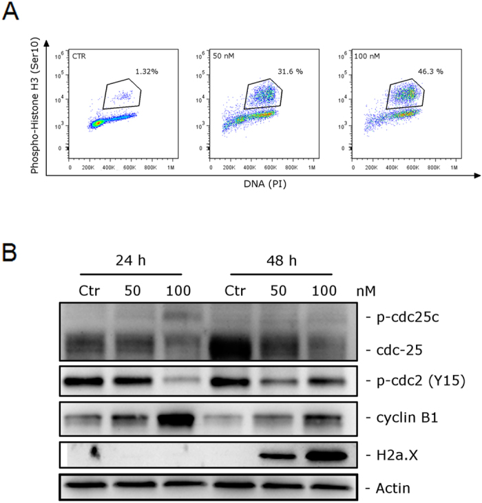
(A) Representative histograms of mitotic cells with phosphorylated histone-H3 after treatment with 4i at the indicated concentrations in HeLa cells. Data are representative of two experiments with similar results. (B) Effect of 4i on cell cycle checkpoint proteins and expression of p-H2A.XSer139. HeLa cells were treated for 24 or 48 h with the indicated concentrations of 4i. The cells were harvested and lysed for detection of the expression of the indicated protein by western blot analysis. To confirm equal protein loading, each membrane was stripped and reprobed with anti-β-actin antibody.