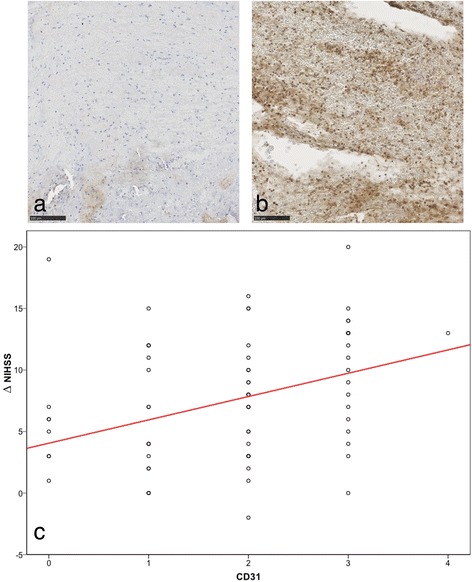
Fig. 2.

Relation between CD31+ cells and early patient improvement. Example of an immunostained specimen with almost no stained nucleated cells (a) and an example with many CD31+ stained cells (b). In this example, there is almost no "staining-negative" cell visible. c Boxplot analysis with regression line in red, showing the relationship between amount of CD31+-cells (0 = no cells, 1 = sporadic, 2 = few, 3 = some, 4 = many) and early patient improvement (represented by ΔNIHSS)
