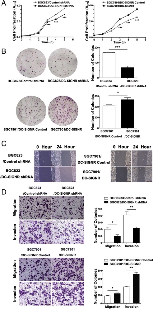
Fig. 3.

The function of DC-SIGNR in vitro. a, cell proliferation was measured by MTT assay. BGC823 cells were infected with sh-DC-SIGNR. SGC7901 cells were infected with DC-SIGNR mediated by lentivirus. b colony-forming growth assays were performed to determine the proliferation of sh-DC-SIGNR infected BGC823 cells and DC-SIGNR mediated by lentivirus infected SGC7901 cells. Colonies were counted and captured. c wound-healing assays were conducted to determine the rate of migration by measuring the distance from one edge of the wound to the other side. d transwell assays were conducted to investigate changes in BGC823 cells and SGC7901 cells migration and invasion. All experiments were performed in biologic triplicates with three technical replicates; *P < 0.05, **P < 0.01 and ***P < 0.001
