
Fig. 1.
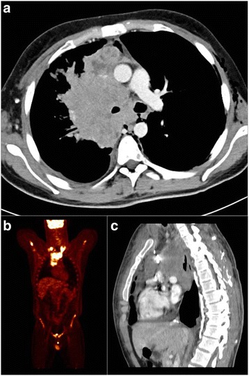
Disease status at diagnosis. Panel a, c: CT scan showing bulky mediastinal mass with involvemente of right lung ilum, compression of the right superior bronchus, and dislocation of the trachea and right intermediate bronchus with pleural and pericardial effusion (panel a axial, panel c sagittal). Panel b: PET scan showing supraclavicular involvement of the disease and absence of liver metastases. CT, computed tomography; PET positron emission tomography