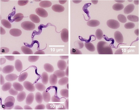
Fig. 2.

Light micrographs of Giemsa-stained blood smears from camel samples. a Trypanosoma evansi with a small subterminal kinetoplast at the pointed posterior end, a long free flagellum and a well-developed undulating membrane. b Trypanosoma vivax with a long free flagellum, an inconspicuous undulating membrane, a rounded posterior end and a large terminal kinetoplast. c Mixed infection. Scale-bars: 10 μm
