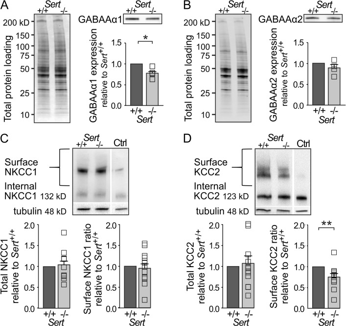
Figure 5.
Reduced inhibitory drive in Sert−/− rats is associated with a decrease in GABAA α1 subunit expression as well as a reduced KCC2 chloride extruder protein expression within S1. (A) Western blot analysis of GABAA α1 subunit expression in S1 total protein lysates of Sert+/+ (n = 4) and Sert−/− rats (n = 4). Left, loading control. Right, western blot and histogram showing total GABAA α1 receptor subunit expression normalized to Sert+/+. (B) Western blot analysis of GABAA α2 receptor subunit expression in S1 total protein lysates of Sert+/+ (n = 4) and Sert−/− rats (n = 4). Left, loading control. Right, western blot and histogram showing total GABAA α2 subunit expression normalized to Sert+/+. (C) Left, surface and internal NKCC1 protein levels were determined by BS3 cross-linking method using S1 protein lysates of Sert+/+ and Sert−/− rats with non-crosslinked control (Ctrl). Right, histograms showing quantification of total NKCC1 expression normalized to γ-tubulin and surface NKCC1 expression normalized to internal expression in Sert+/+ (n = 12) and Sert−/− (n = 12) rats. (D) Left, surface and internal KCC2 protein levels were determined by BS3 cross-linking method using S1 protein lysates of Sert+/+ and Sert−/− rats with non-crosslinked control (Ctrl). Right, histograms showing quantification of total KCC2 expression normalized to γ-tubulin and surface KCC2 expression normalized to internal expression in Sert+/+ (n = 12) and Sert−/− rats (n = 12). All data are presented as mean ± SEM, *P < 0.05 and **P < 0.01 (t-test).

