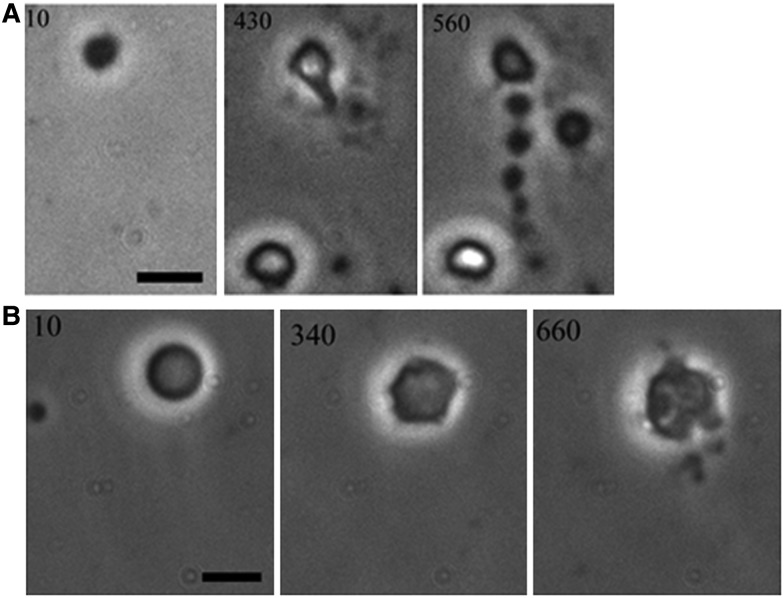
Figure 1. Examples of proliferative events in L-forms of B. subtilis, as viewed by phase contrast microscopy.
Numbers refer to time (min) of observation (from ref. [19]). (A) An event we called extrusion–resolution. A spherical L-form increases in size, then a tubular protrusion emerges which breaks down into a chain of connected progeny cells. (B) A larger L-form again starts as a sphere, then undergoes pulsating changes in shape before multiple small progeny cells erupt from at least three different places on the cell surface.

