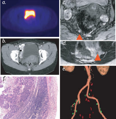
Figure 3. Pelvic Nodal Staging.
Nodal staging in patient with colorectal cancer. A PET scan using 18FDG as a tracer (A) and a CT scan (B) were interpreted as negative for nodal metastases. LMRI identified six small pelvic lymph nodes ([C] and [D]; red arrowheads), which had magnetic parameters of malignancy. Semiautomated reconstruction (E) identifies multisegmental metastases, subsequently proven histologically (F). For 3D reconstruction of pelvic nodal anatomy see Video 1.

