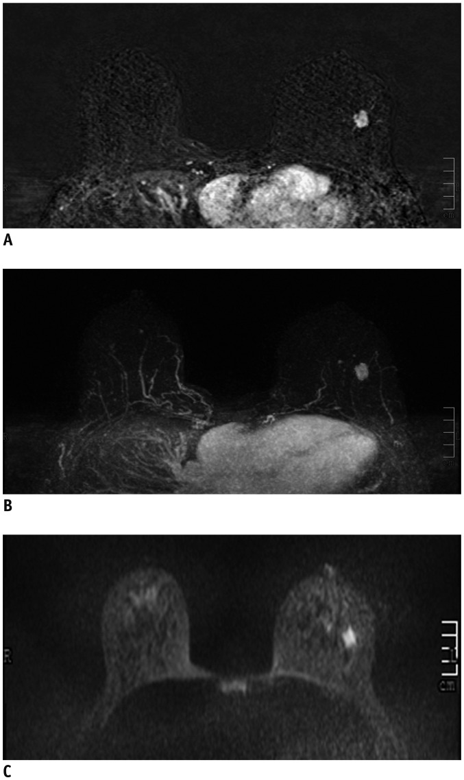
Fig. 1. 40-year-old woman with dense breast tissue.
FAST (A) and MIP (B) of MRI show 11-mm homogeneously enhancing mass with circumscribed margins in left breast, which was classified as probably benign (BI-RADS 3). However, DWI (C) shows high signal (low signal on apparent diffusion coefficient), which was classified as malignant (BI-RADS 4), and biopsy yielded invasive ductal carcinoma.
BI-RADS = Breast Imaging-Reporting and Data System, DWI = diffusion-weighted imaging, FAST = first post-contrast subtracted, MIP = maximum-intensity projection

