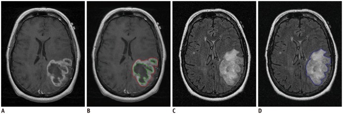
Fig. 1. MR images of example TCGA GBM case (TCGA-06-0213, 55-year-old female patient).
Tumor segmentation was performed semi-automatically with TumorPrism3D.
A. T1W post-contrast image. B. Segmented ROIs for enhancement (red) and necrosis (green) components. C. FLAIR image. D. Segmented ROI for non-enhancing T2 high signal intensity component (blue). FLAIR = fluid-attenuated inversion recovery, GBM = glioblastoma multiforme, ROI = region of interest, TCGA = The Cancer Genome Atlas, T1W = T1-weighted

