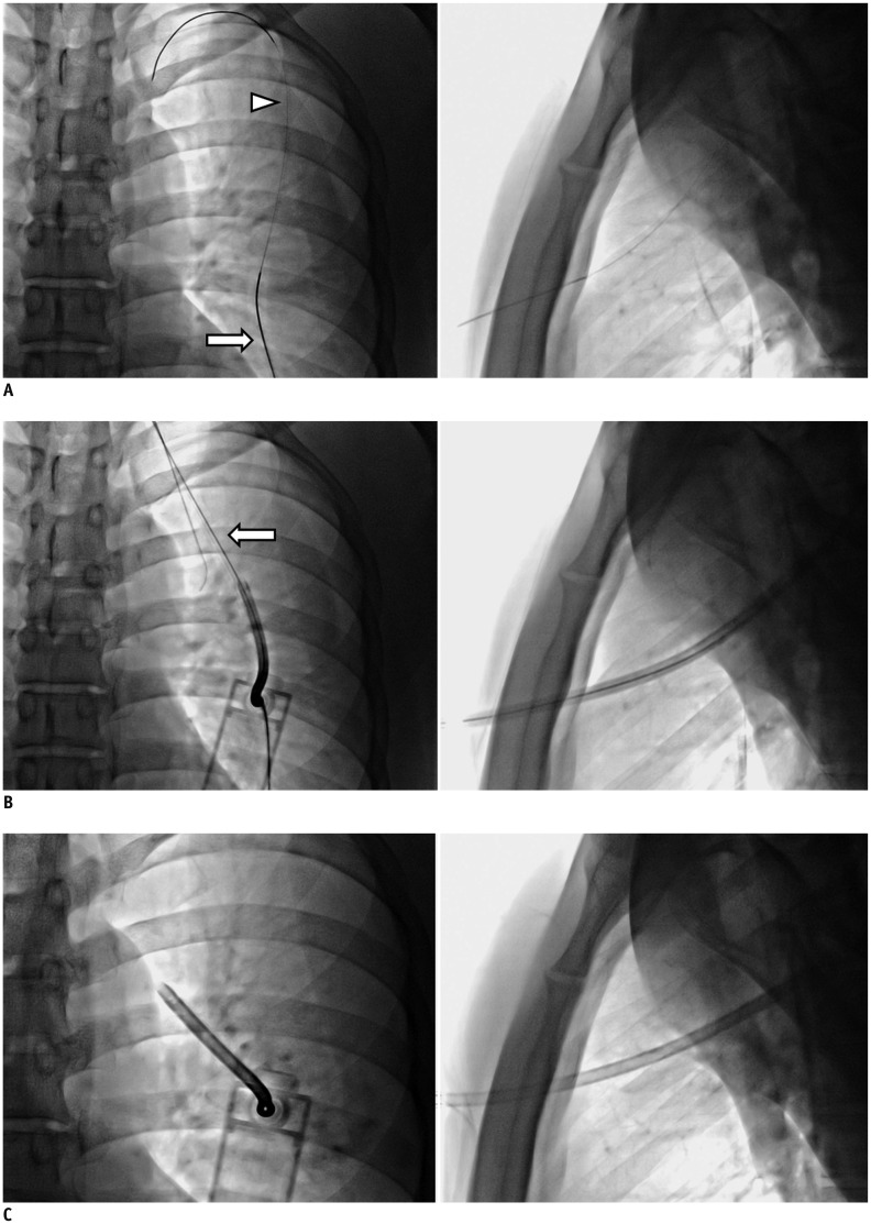
Fig. 2. Thoracic vent insertion under fluoroscopic guidance.
A. 21-gauge Chiba needle (arrow) is inserted into second to fourth intercostal space in midclavicular line. Following confirmation of needle position within pleural space under bi-plane fluoroscopic guidance, 0.018-inch hair-wire (arrowhead) and yellow sheath are introduced sequentially. B, C. Guide wire is then exchanged for 0.035-inch radiofocus stiff wire (arrow), and thoracic vent device is inserted over-wire with 7-Fr dilator.

