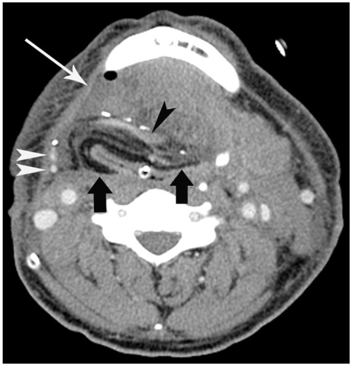
Fig. 4. 55-year-old man with flap success.
Patient underwent reconstruction by radial forearm flap after resection of laryngeal cancer. Contrast-enhanced CT scan 7 days after operation shows fat-containing flap (arrows) and small peri-flap fluid collection (long arrow) with small air bubble in right submandibular space. Note enhanced vascular pedicles (black and white arrowheads) of flap.

