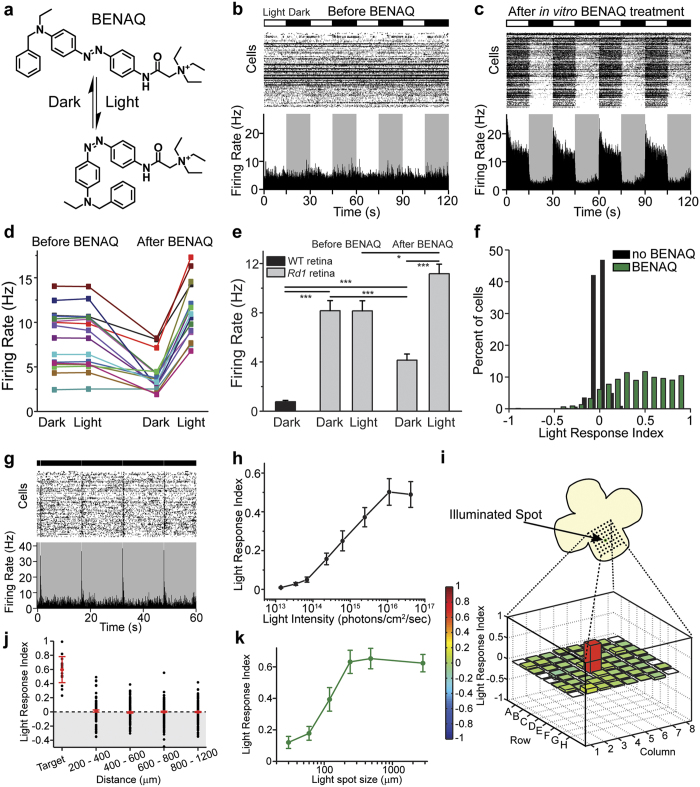
Figure 1. BENAQ is a photoswitch that restores spatially precise light responses to the blind retina.
(a) Structure of BENAQ. Visible light converts BENAQ from the trans to the cis form and then the compound quickly relaxes back to trans in the dark. (b,c) MEA recordings from an rd1 mouse retina before (b) and after (c) BENAQ treatment. Raster plots of individual RGC activity and average firing rate plots are shown. Alternating light (white) and dark (black) intervals plotted at the top. (d,e) Average rd1 retinal firing rate in the dark and light before (left) and after (right) BENAQ treatment (n = 16 retinas). Average WT retinal firing rate in the dark (e, left) (n = 8 retinas). Data are mean ± SEM. (f) LRI value distributions for RGCs from untreated (black) (median LRI = 0.00) and BENAQ-treated (green) rd1 retinas (median LRI = 0.51, p < 0.001, rank sum test). (g) MEA recording of a BENAQ treated rd1 mouse retina stimulated with 100 ms white light flashes every 15 seconds. (h) White light intensity – response curve for BENAQ treated rd1 retinas (n = 5 retinas). Light intensity threshold for driving RGC activity = 7 × 10¹³ photons/cm2/sec. Data are mean ± SEM, n = 5 retinas. (i) Rd1 retinal light response to targeted illumination of electrode E4 with a 120 μm-diameter light spot. Only electrode E4 (red) recorded an increase in RGC activity in response to white light (bottom). LRI values are color-coded (scale at left) and also represented by bar height. (j) Targeted illumination elicits an increase in activity in stimulated RGCs and has no effect on surrounding RGCs (n = 17 cells and n = 903 cells, respectively, from seven retinas). LRI values of RGCs (black circles) as a function of distance from the target electrode, displayed in 200 μm bins. Median plus and minus the 95% confidence intervals are shown in red. See also Supplementary Table S1. (k) Responses of BENAQ-treated rd1 RGCs to stimulation with light spots of increasing diameter. The light response saturates at 240 μm-diameter spot size. Data are mean ± SEM; n = 20 cells.

