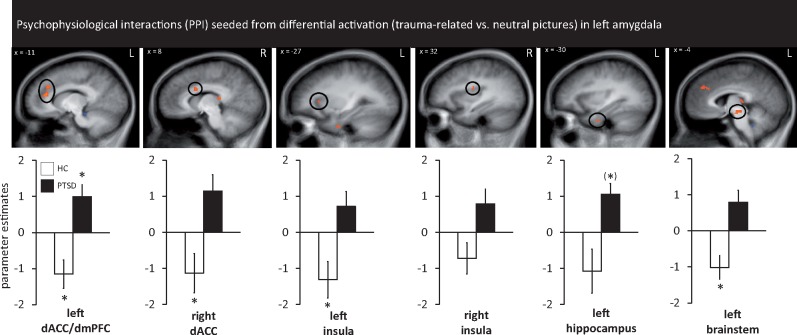
Fig. 4.
PPIs seeded from differential activation (trauma-related vs neutral pictures) in left amygdala (as shown in Figure 3). Regions showing higher PPI connectivity (all P <0.050 corrected) between patients suffering from PTSD and HC with the seed region in left amygdala: significant differences were found in left dorsal ACC/dorsal medial prefrontal cortex (mPFC) for both groups, right dorsal ACC for HC, left insula for HC, left hippocampus (marginally significant for PTSD) and left brainstem (HC). Asterisks mark significance against baseline.

