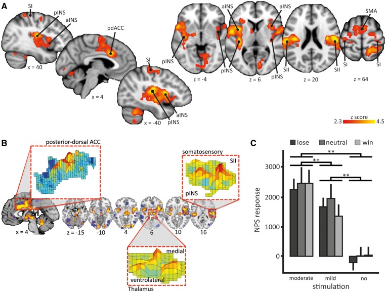
Fig. 3.
Neurological pain signature response and brain correlates of moderate compared to mild pain. (A) An univariate analysis showed that moderate pain was associated with higher brain activations compared to mild pain (contrast: [win_mod + neut_mod + lose_mod] > [win_mild + neut_mild + lose_mild]): anterior insula (aINS), posterior insula (pINS), primary somatosensory cortex (SI), secondary somatosensory cortex (SII), posterior-dorsal anterior cingulate cortex (pdACC) and supplementary motor area (SMA). Images are displayed in neurological convention, i.e. right side of the brain is on the right. Coordinates are given in MNI space. Statistical inference was based on a voxel-based threshold of z = 2.3, cluster corrected at p <.05 on a whole brain level. For details see Table 1; (B) A priori defined pattern of the Neurological Pain Signature (NPS). The inset show examples of the pattern distribution of voxel weights within certain brain areas; (C) NPS responses in the winning, losing, and neutral condition with mildly painful, moderately painful, or no stimulation of the wheel of fortune game; mean scalar values expressing the NPS across subjects; error bars: standard error of the mean. Post hoc comparisons ** P < 0.01. Scaling of the NPS values depends on many factors such as voxel size, contrast weight, field strength, etc. Because only a within-study comparison was of interest here, we did not attempt to equate scaling of the NPS values with previous studies.

