. 2016 Oct 21;8(2):1450–1453. doi: 10.1039/c6sc03702j

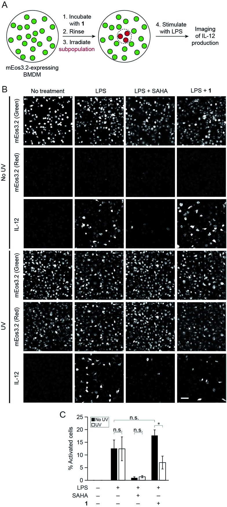
Fig. 4. Photo-uncaging of 1 is spatially selective and leads to a decrease in IL-12 production by a subpopulation of macrophages. BMDM expressing mEos3.2 were pre-treated with 1 (3 μM), and the center of the dish was irradiated with UV light (epifluorescence using DAPI filter, 30 s). The BMDM were then activated with the indicated combinations of LPS (100 ng mL–1) and/or SAHA (500 nM). After 6 h, the cells were fixed, and immunofluorescence labeling for IL-12 was performed. (A) Schematic of experimental setup. (B) Maximum intensity z-projection images of cells in the periphery (top three rows, no UV) and center (bottom three rows, UV) of the same dish, imaged by confocal microscopy. Green indicates non-photoconverted state of mEos3.2 and red indicates photoconverted state of mEos3.2. Scale bar: 50 μm. (C) Quantification of data from (B), where % activated cells represents the percentage of IL-12-positive cells. n.s. = not significant, *p < 0.05.

Fig. 4