
Figure 3.
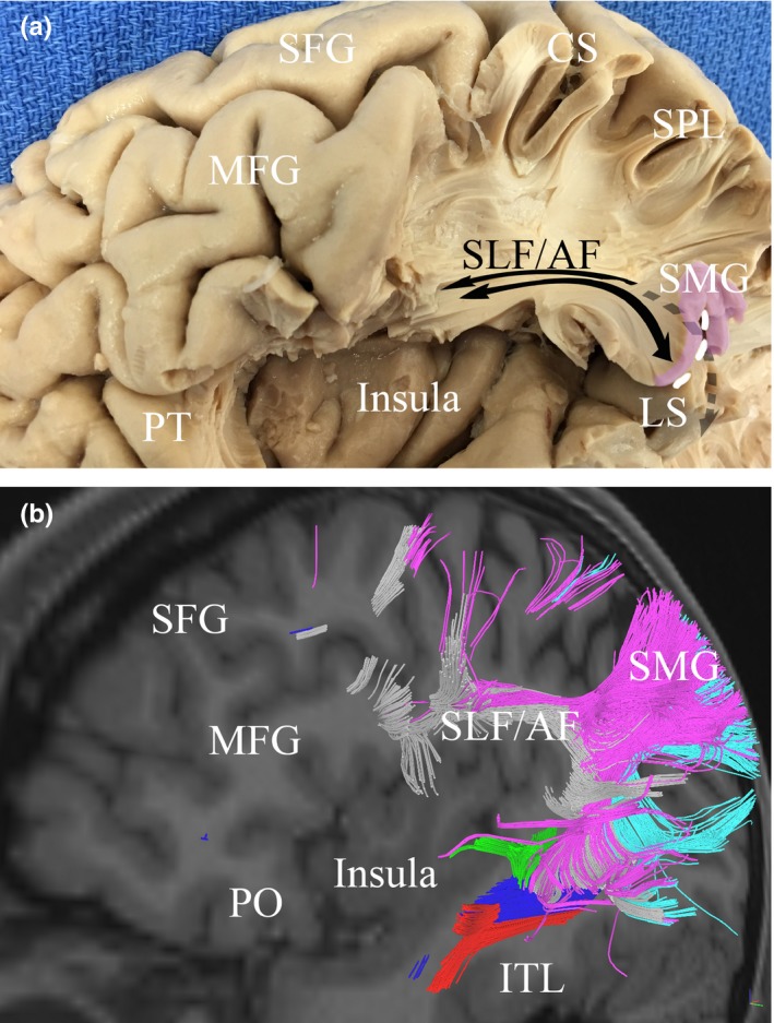
The relationship between the inferior parietal lobule (IPL) and the insula. Gross dissection (a) and tractography (b) illustrate the proximity of the insula to fibers connecting the IPL to the frontal lobe. Fibers connecting to frontal regions and the AG curve with the arcuate fasciculus around the posterior edge of the insula before becoming part of the superior longitudinal fasciculus (SLF), passing directly superior to the insula. Similarly, fibers connecting to frontal regions and the SMG join the SLF to course antero‐posteriorly just above the insula. Red, inferior longitudinal fasciculus; Blue, inferior fronto‐occipital fasciculus; Green, middle longitudinal fasciculus; Purple, SMG/fibers connecting to the SMG; Light Blue, AG /fibers connecting to the AG; white, superior longitudinal fasciculus; CS, central sulcus; ITG, inferior temporal gyrus; LS, lateral sulcus; MFG, middle frontal gyrus; PO, pars orbitalis; SFG, superior frontal gyrus; SLF/AG, superior longitudinal fasciculus/arcuate fasciculus; SMG, supramarginal gyrus