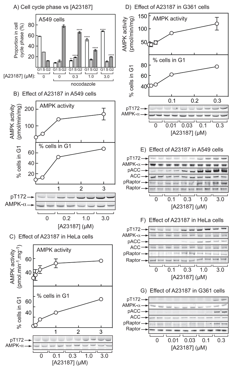
Figure 2. Effect of A23187 on AMPK activation and cell cycle arrest in A549, HeLa and G361 cells.
(A) A549 cells were treated with the indicated concentration of A23187 or vehicle (DMSO) for 20 hr with (where indicated) nocodazole (70 ng.ml-1) added for a further 18 hrs. The cells were then fixed, stained with propidium iodide and DNA content analyzed by flow cytometry. Bars show the percentages of cells with G1, S and G2/M phase DNA content (mean ± SD, n = 3); significant differences in the % of cells in the same cell cycle phase compared with DMSO control by 2-way ANOVA are shown: **p<0.01, ****p<0.0001. Similar results were obtained in three identical experiments. (B) AMPK activity measured in immunoprecipitates (top), percentage of cells in G1 phase (middle) (both mean ± SEM (n = 3)), and Thr172 phosphorylation (bottom) in A549 cells treated with various concentrations of A23187 for 20 hr;. (C) As (B), but in HeLa cells. (D) As (B), but in G361 cells (note different scale on x axis). (E) Phosphorylation of AMPK (Thr172), ACC (Ser79) and Raptor (Ser792) in A549 cells incubated as in (B). (F) As (E), but in HeLa cells incubated as in (C). (G) As (E), but in G361 cells incubated as in (D).

