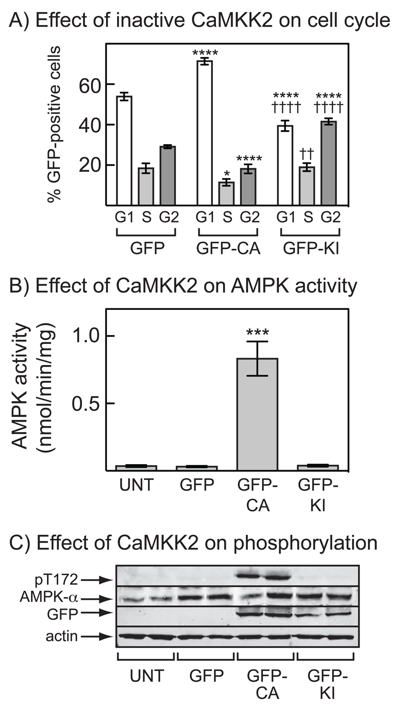
Figure 4. Cell cycle arrest and AMPK activation in G361 cells requires the kinase activity of CaMKKβ.
(A) Percentage of cells in G1, S or G2/M for cells transfected with DNAs encoding GFP, GFP-CA or GFP-KI (mean ± SD, n = 3). After 36 hr, cells were treated with nocodazole (70 ng.ml-1) and 18 hrs later were fixed, stained with propidium iodide and the DNA content analyzed by flow cytometry. The cytometer was set-up to analyze only cells expressing GFP. Significant differences by 2-way ANOVA compared with values for the same cell cycle phase with GFP alone (*p<0.05, ****p<0.001) or between GFP-CA and GFP-KI (††p<0.01, ††††p<0.0001) are shown. Similar results were obtained in two identical experiments. (B) AMPK activity measured in immunoprecipitates from untransfected cells or cells transfected with DNAs encoding GFP, GFP-CA or GFP-KI. Results are mean ± S.D. (n = 2); significant differences compared with untransfected cells are shown: ***p<0.001 (1-way ANOVA). (C) Analysis by Western blotting of extracts of the cells shown in (B) (n = 2).

