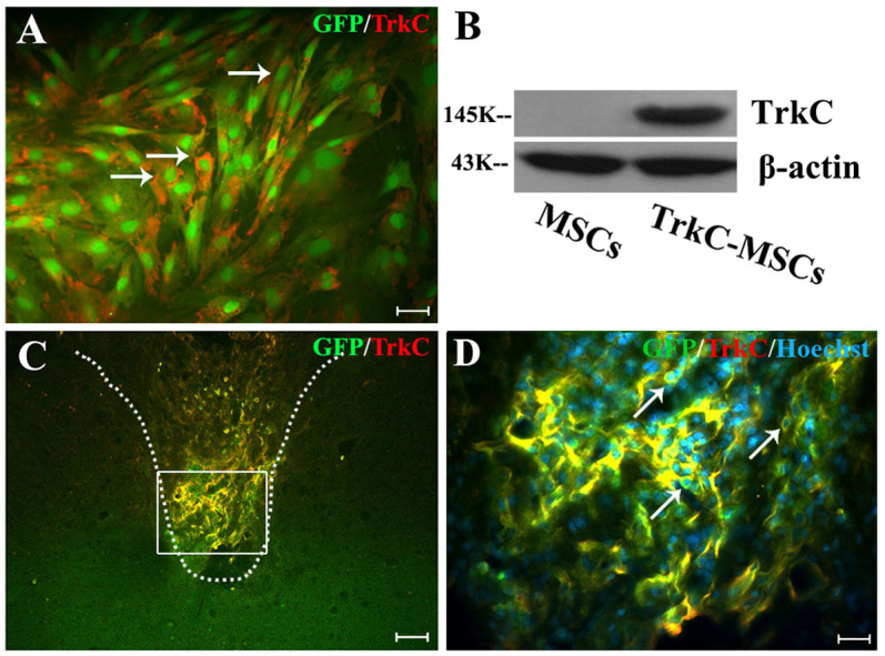
Figure 1. In vitro and in vivo analysis of adenoviral (Ad) vector-mediated transgene expression.

(A) Performing TrkC immunofluorescence staining, 48 h after infection GFP-MSCs with Ad-TrkC. More than 80% cultured GFP-MSCs (green) expressed the TrkC gene product (red, arrows). Scale bar: 20 μm. (B) Transgenic MSCs were analyzed for the presence of TrkC using Western blot, 48 h after Ad vector transduction. Ad-TrkC transduced MSCs expressed TrkC protein, but TrkC protein could not be detected in non-transduced MSCs. Gels/blots were run under the same experimental conditions and β-actin was shown as a control. The cropped blots images were shown in the full-length blots are presented in Supplementary Figure 1. (C) In vivo confirmation of Ad vector-mediated TrkC expression in the GFP-MSCs (yellow) at 30 d after EB injection. (D) Showing higher magnification of GFP/TrkC/Hoechst33342 positive MSCs (yellow, arrows) in the rectangle boxes of (C). Scale bars: (C) = 80 μm; (D) = 20 μm.
