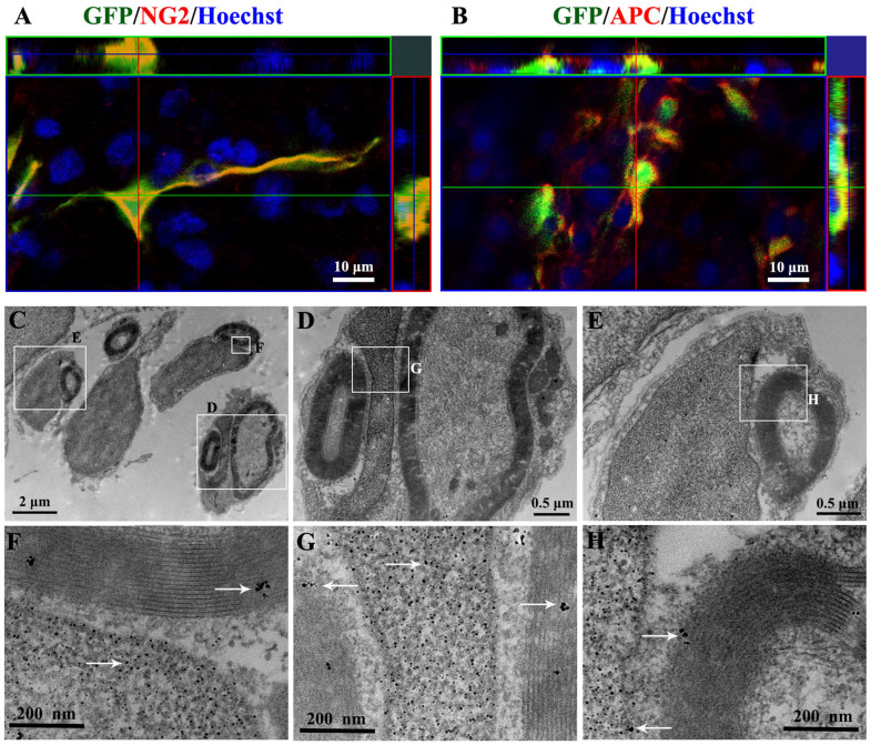
Figure 6. Confocal microscope and immunoelectron microscope images showed the differentiation of grafted MSCs into the oligodendrocyte-like cells within the demyelination site of spinal cord in the TrkC-MSCs+EA group.
(A) The confocal imaging confirmed the colocalization of NG2 expression (red) and GFP positive MSCs (green) grafted. (B) The confocal imaging confirmed the colocalization of APC expression (red) and GFP positive MSCs (green) grafted. (C) Showing four GFP positive cells giving rise to the myelin profiles encircling an axon respectively. (D, E) Showing the higher magnification of the rectangle boxes in (C). Some GFP reaction products (gold-particles, white arrows) exist in the nucleus, myelin and cytoplasm of the grafted cells ((F, G and H) showing higher magnification of the rectangle boxes in (C–E)).

