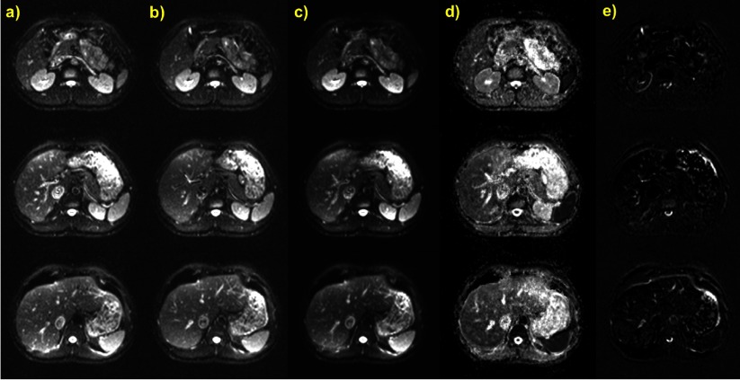
Figure 4.
Three abdominal slices of a DW-EPI of a healthy volunteer acquired during multiple ABC breath holding with the same settings at two time points (a) and (b) during the same session (b = 0 s mm−2, identical slice position) to demonstrate averaging with ABC breath holds. (c) is the average of (a) and (b). (d) ADC maps produced by aggregating the DW images from the time points (a) and (b) at each slice position (b = 0, 100, 500, 750 s mm−2). (e) Difference images (b) − (a). No image registration was used in any operation and displayed windowing is the same in all cases except for the ADC maps.

