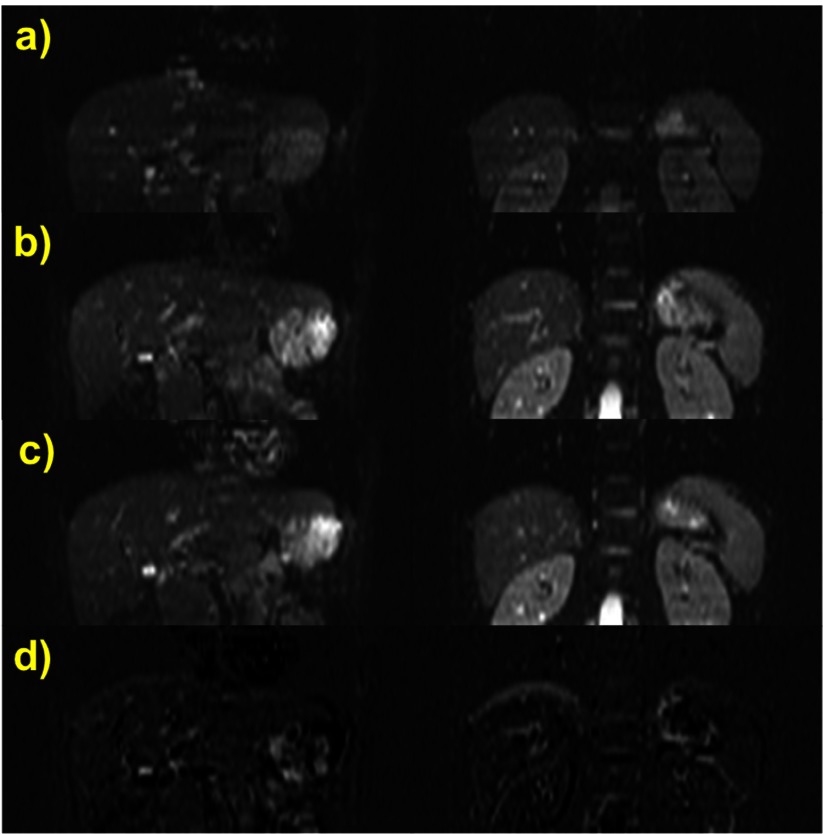
Figure 5.
Two coronally reformatted slices of a DW-EPI (b = 0 s mm−2) of a healthy volunteer acquired during (a) standard operator-instructed self-induced multiple breath holds; (b) ABC-controlled multiple breath holding and (c) repetition of (b). (d) Difference images (b) − (c), demonstrating good intra-session registration using the ABC. No image registration was used and the slice location and windowing is identical for all displayed images.

