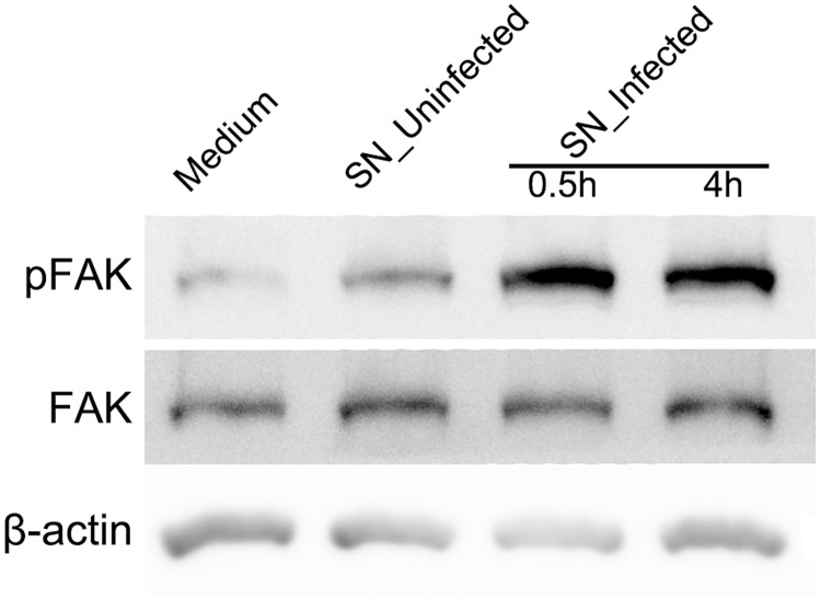
Fig 4. FAK is activated by conditioned media from infected monocytes.
Representative Western blots of anti-pFAK, anti-FAK and anti-β-actin obtained with RPE cell lysates after treatment of conditioned media. RPE cells were incubated with standard medium, supernatants from uninfected THP-1 cells (SN_Uninfected) for 0.5 h, or supernatants from infected THP-1 cells (SN_Infected) for 0.5 and 4 h. β-actin served as loading control. Figures were selected as representative data from three independent experiments.

