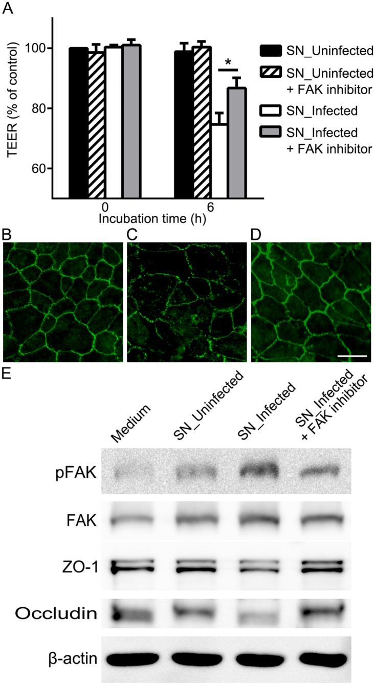
Fig 5. Inhibition of FAK signaling attenuates the disruption of outer BRB.

(A) Transepithelial electrical resistance was measured before the treatment (0 h) and 6 h after the treatment of supernatants from THP-1 cells that were not infected (SN_Uninfected) or from THP-1 cells infected with T. gondii (SN_Infected) with or without FAK inhibitor (PF-573228, 1μM). (B-D) Expression of ZO-1 was evaluated by immunocytochemical staining of ZO-1 (green) 6 h after the treatment of supernatants from uninfected THP-1 cells with FAK inhibitor (B), supernatants from infected THP-1 cells without FAK inhibitor (C) or with FAK inhibitor (D). (E) Representative Western blots of anti-pFAK, anti-FAK, anti-ZO-1, anti-occludin and anti-β-actin obtained with RPE cell lysates after treatment of conditioned media with or without FAK inhibitor for 6 h. β-actin served as loading control. Data were presented as the mean ± SEM of five independent experiments. Figures were selected as representative data from three independent experiments. *, P<0.05. Scale bar = 20 μm.
