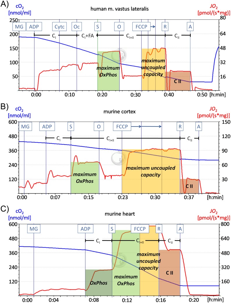
Fig 1. Representative high-resolution respirometry recordings of the human vastus lateralis muscle and the murine prefrontal cortex and heart.
Representative measurements of high-resolution respirometry recordings are shown for the human vastus lateralis muscle (A), the murine prefrontal cortex (B) and the murine heart (C). The blue line represents the oxygen concentration in the chamber, while the red line indicates the oxygen flux. (A) For experiments with human vastus lateralis muscle samples 10 mM pyruvate were added before the recording started. After administration of 5 mM malate (M), 10 mM glutamate (G) and 5 mM ADP complex I activity (CI) was recorded and the leak respiration was calculated (respiration after MG/respiration after ADP). 10 μM cytochrome c was added to survey the integrity of the outer mitochondrial membrane. 1 mM octanoyl carnitine was administered to measure the fatty acid induced respiration. Further addition of 10 mM succinate, which is the complex II substrate, enables the determination of the maximum OxPhos capacity. The ATP synthase inhibitor oligomycin (O) (5 μM), which was not used for the murine heart (C) and soleus muscle was administered. After adding the uncoupling agent carbonyl cyanide p-(trifluoromethoxy)-phenylhydrazone (0.5, 0.25, 0.25 μM FCCP) the maximum uncoupled capacity was measured. Finally complex I was inhibited by rotenone (R) (0.5 μM) allowing the determination of complex II activity (CII). In the end complex III was inhibited by administration of antimycin A (5 μM). (B) The protocol for murine cortex and liver matched the human muscle protocol with the following exceptions: cytochrome c was added after pyruvate before the recording started and octanoyl carnitine was not used. (C) The protocol for murine heart and soleus muscle is equivalent to the one used for murine cortex (B) except for the oligomycin administration.

