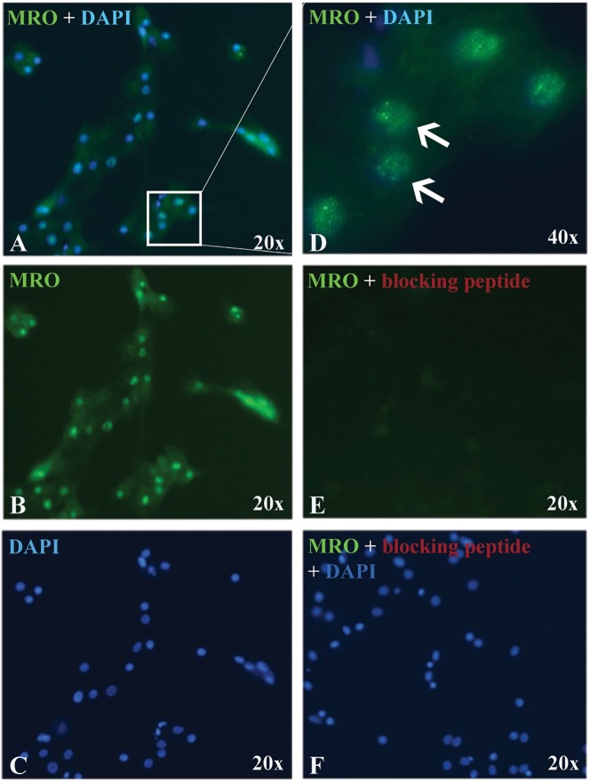
Fig 4. MRO expression in GCs.
Representative micrograph of GCs immunostained with MRO-FIL antibody (0.4 ug/ml) and counterstained with DAPI (nuclei). (A) Merged signals, (B) MRO only, (C) DAPI only. (D) Merged signals under high magnification (x40) micrograph showing MRO staining in the nucleus, as well as in the cytoplasm. Antibody specificity was validated with (E) MRO blocking peptide (CAT# ab206335). (F) Counterstain with DAPI. All images are taken with the same exposure time.

