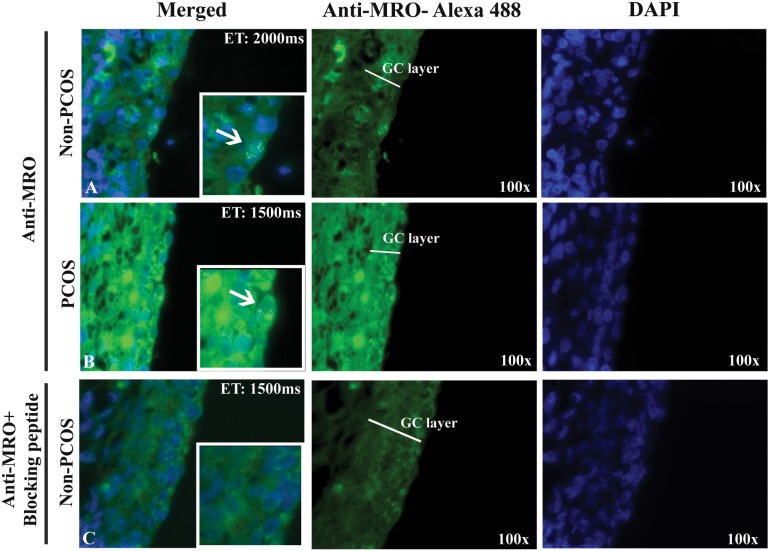
Fig 6. MRO expression in human ovarian sections.
Immunofluorescence was performed on non-PCOS (A) and PCOS (B and C) ovarian sections using the FL-248 antibody (4ug/ml). Nuclear staining is apparent in the granulosa cells (magnified images), while the blocking peptide abolished the MRO signal (C). MRO expression was observed in green (Alexa-fluor 488) and nucleus in blue (DAPI). Magnification: x100. Duration of exposure: 2000 milliseconds (non-PCOS) and 1500 milliseconds (PCOS) demonstrating that even with short exposure the fluorescence intensity is higher in PCOS samples.

