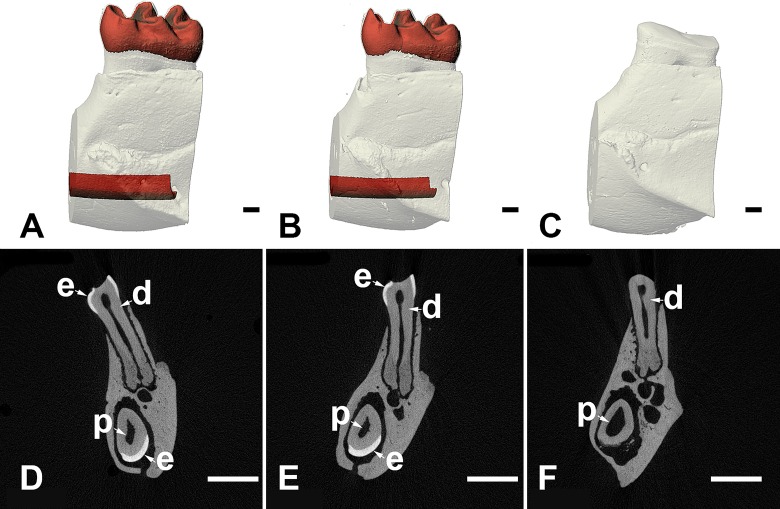
Fig 5. μCT evaluation showed Slc13a5-/- mice lacked mature, densely-mineralized enamel in the molars and incisors.
Representative μCT images of volumetric renderings (A-C) or slices (D-F) of first molars and incisors from 32-week-old WT (A, D), Slc13a5+/- (B, E) and Slc13a5-/- (C, F) mice. Tissue colored in red in panels A and B corresponds to mature enamel in molars or incisors. Tissue colored white in panels A to C corresponds to bone or dentin, both of which have generally similar radio-density. Enamel (e), dentin (d), and pulp (p) are indicated in molars (top) and incisors (bottom) in panels D through E. Size bar in panels A-C = 100 micrometers; size bar in panels D-E = 1 millimeter.

