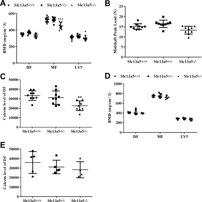
Fig 7. Measurements of bone mineral density, strength and formation.
At 13 weeks old, (A) Slc13a5-/- mice had similar BMD in distal femur (DF) and 5th lumbar vertebrae (LV5) to Slc13a5+/+ mice, but had decreased BMD in mid femur (MF), and a trend with decreased bone strength (B) (P = 0.096), and decreased calcein incorporation (C) compared with Slc13a5+/+ (n = 9 each group). At 32 weeks old, Slc13a5-/- mice had similar BMD in DF, LV5 and MF (D) and calcein incorporation (E) to Slc13a5+/+ (n = 5 each group). ** P < 0.01; *** P < 0.001 vs age-matched Slc13a5+/+ mice.

