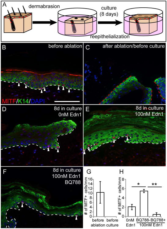
Figure 4. Edn1 promotes generation of epidermal melanocytes in human scalp tissue.
(A) Schematic figure of human scalp explant culture experiment. (B-F) Immunohistochemical analysis with K14 and MITF of sections of human scalp before ablation (B), after ablation of skin prior to culture (C), 8 days in culture in the absence (D) and presence Edn1 (E) and presence of both Edn1 and BQ788 (F). (G and H) Quantification of the number of MITF+ cells in human scalp before culture (G) and after 8 days in culture (H). White dashed line indicates the border between inter follicular epidermis and dermis. Red dashed line indicates the border between hair follicle epidermis and dermis. Arrowheads indicate MITF+ cells. Data are presented as the mean ± SD. *, p<0.05, **, p<0.005. Scale bar, 100μm.

