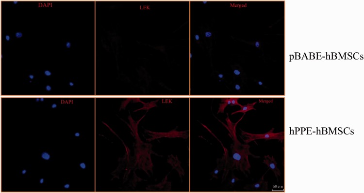
Figure 3.
The different expressions levels of Leu-enkephalin (L-EK) protein were compared between the pBABE-hBMSC group and the hPPE-hBMSC group by immunofluorescence. pBABE, a retroviral vector; hPPE, human proenkephalin; hBMSCs, human bone marrow stem cells. pBABE-hBMSCs, the pBABE-hBMSCs group; hPPE-hBMSCs, the hPPE-hBMSCs group. Blue fluorescence marks the nucleus of the hBMSCs by 4′6-diamidino-2-phenylindole. Red fluorescence marks L-EK protein. Double-labeled cells, with a blue fluorescent nucleus and a red cytoplasm, represented hBMSCs that expressed the L-EK. There were no differences in nuclear staining between the groups. Little L-EK expression was observed in the pBABE-hBMSC group, whereas greater expression was detected in the hPPE-hBMSC group. All images were obtained on a laser scanning confocal microscope (Leica). Scale bars = 50µm. n = 6.

