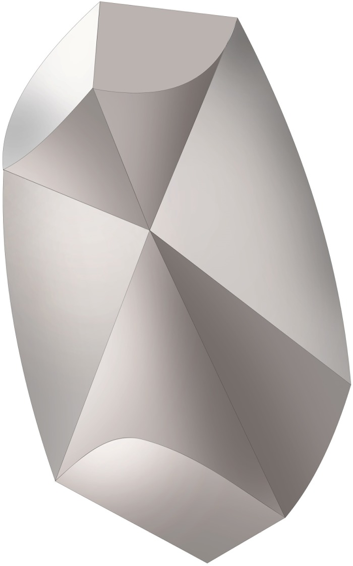
Fig 6. 3 D-model of the inner structure of a single human otoconium.
Differences in size among the rhombohedral faces clearly correlate with variations in volume among the 3+3 branches. The inner structure deviates from centrosymmetry indicating non-centrosymmetric mass distribution parallel to the long axis of the otoconium.

