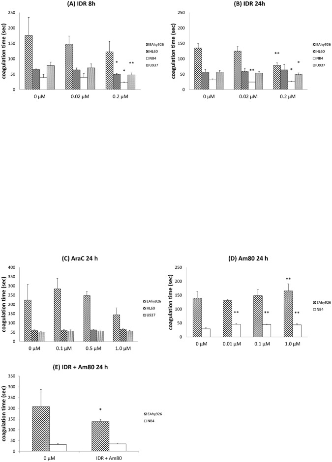
Fig 1. Effects of IDR, AraC, and Am80 on cell surface PCA.
Each cell line (EAhy926, HL60, NB4, and U937) was treated with IDR, AraC, or Am80 at 37°C for 8 or 24 h. (A) IDR (8 h), (B) IDR (24 h), (C) AraC (24 h), (D) Am80 (24 h), and (E) IDR + Am80. PCA was measured by normal plasma-based recalcification time. Data are the mean ± SD (n = 6). Significant differences are indicated by (*) when p < 0.05 compared with 0 μM, whereas (**) indicates a significant difference with p < 0.01 compared with 0 μM.

