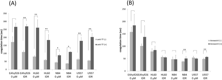
Fig 7. Effects of IDR on cell surface PCA blocked by an anti-TF antibody or annexin V.
(A) To investigate the effect of cell surface TF on PCA induced by 0.2 μM IDR, cell lines were treated with 0.2 μM IDR for 8 h (HL60 cells) or 24 h (EAhy926, NB4, and U937 cells). The cells were then treated with 10 μg/mL mouse monoclonal anti-human TF antibody or the same amount of irrelevant IgG in PBS for 60 min on ice. After washing with PBS, cell surface PCA was assessed as described in Fig 1. Significant differences are indicated by (*) when p < 0.05, whereas (**) indicates a significant difference with p < 0.01. (B) To investigate the effect of cell surface PS exposure in response to 0.2 μM IDR stimulating PCA, after 8 h (HL60 cells) or 24 h (EAhy926, NB4, and U937 cells) of treatment, the cells were treated with 1 μg/mL annexin V in 300 μL annexin V binding buffer at 37°C for 30 min. After incubation, cell surface PCA was assessed as described in Fig 1. Data are the mean ± SD (n = 6). Significant differences are indicated by (*) when p < 0.05, whereas (**) indicates a significant difference with p < 0.01.

