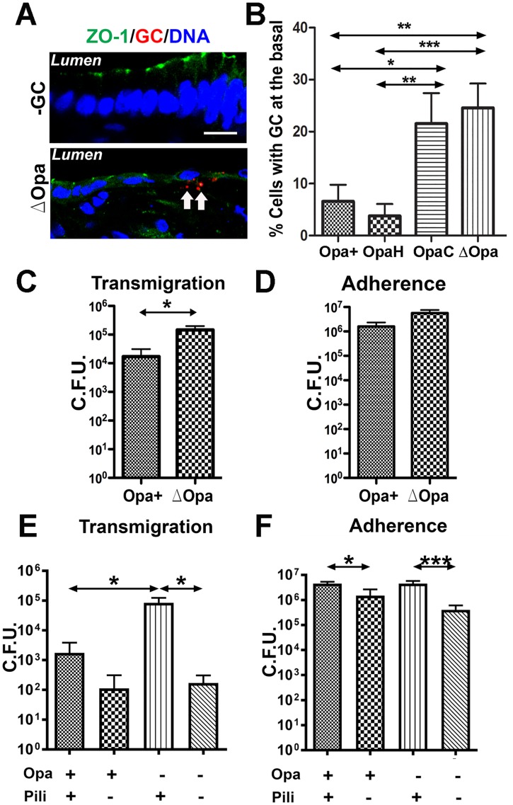
Fig 3. Effects of Opa and pili on GC adherence to and transmigration across polarized epithelial cells.
(A-B) Human endocervical tissue explants were incubated with piliated MS11Opa+, ΔOpa, OpaH, or OpaC for 24 h, stained for ZO-1, nuclei and GC, and analyzed by 3D-CFM (A). GC subepithelial penetration (arrows) was quantified using 3D-CFM images as the percentage of epithelial cells with basal GC staining among the total number of GC-associated epithelial cells (B). Shown are the average values (±SD) of >50 epithelial cells of endocervical tissue explants from two to three human subjects. (C and D) Polarized HEC-1-B cells were apically incubated with piliated MS11Opa+ or ΔOpa. The basal medium was collected after 6 h to determine transmigrated GC (C). The epithelial cells were lysed after 3 h incubation and washing to quantify adherent GC (D). (E-F) Polarized T84 cells were apically incubated with piliated or non-piliated MS11Opa+ or ΔOpa for 6 or 3 h, and the numbers of transmigrated (E) and adherent GC (F) were determined as described above. Shown are the means (±SD) of >6 transwells from 4–6 independent experiments. ***p ≤0.001; **p ≤ 0.01; *p≤0.05.

